Back Of Skull Anatomy : MIDTERM - Anatomy & Physiology Bsc2085 with Ruiz at Miami ... - Learn more about the anatomy and function of the skull in humans and other vertebrates.
Back Of Skull Anatomy : MIDTERM - Anatomy & Physiology Bsc2085 with Ruiz at Miami ... - Learn more about the anatomy and function of the skull in humans and other vertebrates.. Anatomical structures of the skull include: It is comprised of many bones, formed by intramembranous ossification, which are joined together by sutures (fibrous joints). This website is temporarily out of service. Foramina inside the body of humans and other animals. It was then cleaned, adapted and polypainted this model is part of a comparison with the skull of a human.
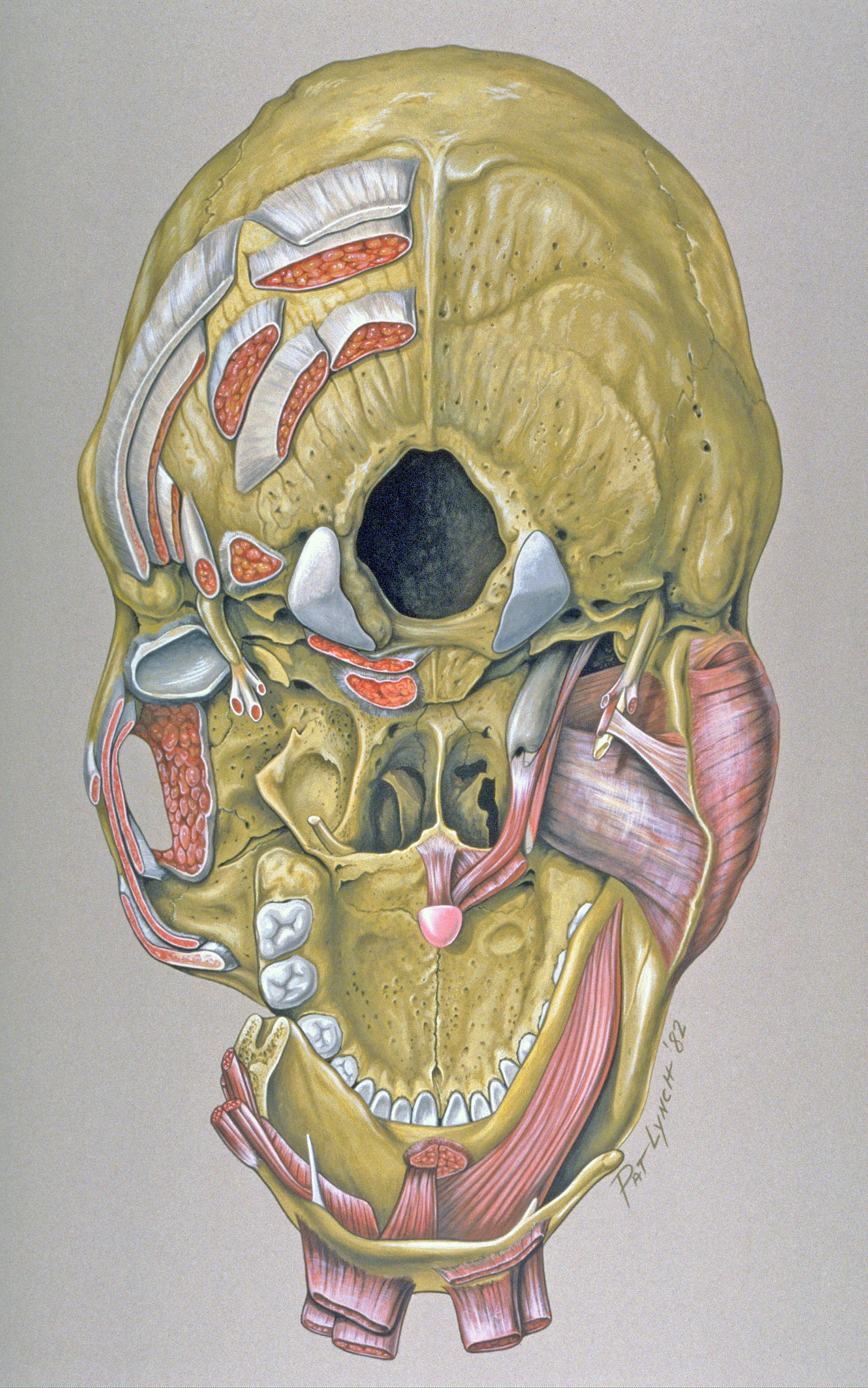
The skull is a bony structure that supports the face and forms a protective cavity for the brain. In order to be light, the skull is made up by flat and irregular bones, and has hollow spaces called the sinuses. The skull base is the inferior portion of the neurocranium. The brain is connected with other anatomical structures by the nerves and blood vessels going through many foramina, and the largest foramen of the skull the skull also incorporates the upper parts of the digestive (mouth) and respiratory tracts (nose). Learn about skull base anatomy with free interactive flashcards.
Human Skull stock illustration. Illustration of front ... from thumbs.dreamstime.com The bbc is not responsible for the content of external websites. The cranium and the mandible. We monitor our sites and will resolve this issue as soon as possible. The skull is the bony skeleton of the head. The major sutures are the coronal suture, sagittal suture, lambdoid suture and squamosal sutures. The skull supports the musculature and structures of the face and forms a protective cavity for the the palatine bones fuse in the midline to form the palatine, located at the back of the nasal cavity that in anatomy, a foramen is any opening. It was then cleaned, adapted and polypainted this model is part of a comparison with the skull of a human. Skull bones aren't fused together at birth.
Cranial cavity , cranial sutures.
Learn about the anatomy of the skull bones and sutures as seen on ct images of the brain. The skull or known as the cranium in the medical world is a bone structure of the head. Excluding ear ossicles, it is made of 22 bones. The skull base is the inferior portion of the neurocranium. The cranium and the mandible. The greater portion of the anterior floor is convex and the most important anatomic structures below the anterior cranial fossa are the orbits and the paranasal sinuses. Skull bones aren't fused together at birth. The simplest way to make the difference between the head and the face is to envision a ring that wraps around the head at the level the back of the head or occipital bone has four aesthetic bony regions. This anatomic region is complex and poses surgical challenges for otolaryngologists and neurosurgeons alike. This view of the skull is dominat. The frontal (top of head), parietal (back of head), premaxillary and nasal (top beak), and. We monitor our sites and will resolve this issue as soon as possible. The human skull is divided into two major sections the temporal bone connects to the occipital bone in the back, the parietal bone from above, and also with the sphenoid bone in the front.
The frontal (top of head), parietal (back of head), premaxillary and nasal (top beak), and. The skull has a single occipital condyle.7 the skull consists of five major bones: It supports and protects the face and the brain. This is a model of the human (homo sapiens) skull. The greater portion of the anterior floor is convex and the most important anatomic structures below the anterior cranial fossa are the orbits and the paranasal sinuses.
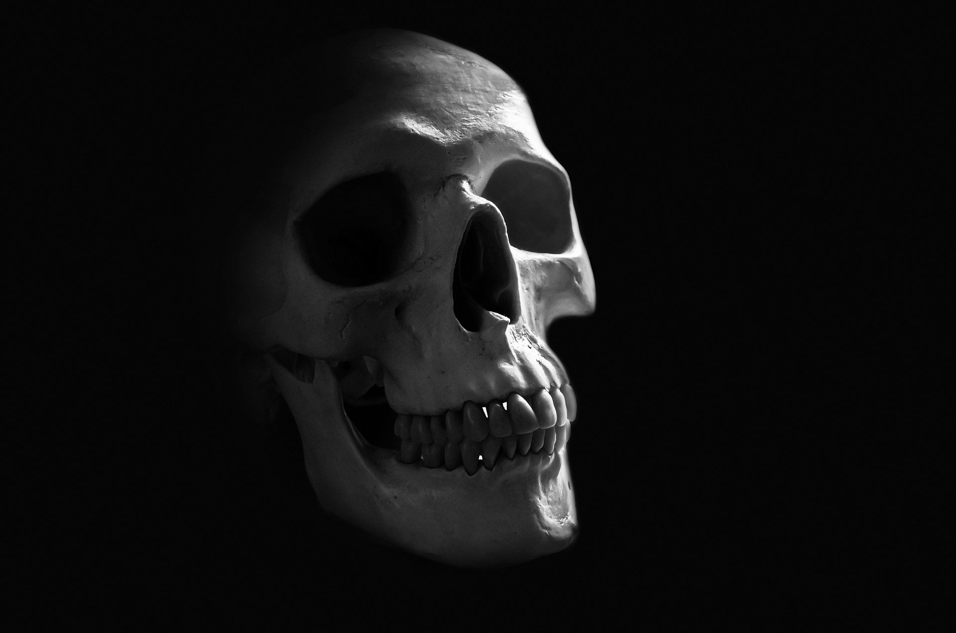
Human Skull HD Wallpaper | Background Image | 1920x1272 ... from images4.alphacoders.com This article describes the anatomy of the skull, including its structure, features, foramina and overview hip and thigh knee and leg ankle and foot nerves and vessels. The skull has a single occipital condyle.7 the skull consists of five major bones: The human skull is divided into two major sections the temporal bone connects to the occipital bone in the back, the parietal bone from above, and also with the sphenoid bone in the front. The skull is the bony skeleton of the head. So, the human skull consists of 23 bones. The bbc is not responsible for the content of external websites. These are the anterior, middle and posterior cranial fossae. Learn more about the anatomy and function of the skull in humans and other vertebrates.
We monitor our sites and will resolve this issue as soon as possible.
Anatomical structures of the skull include: The bbc is not responsible for the content of external websites. Learn skull anatomy with skull bones quizzes and diagram labeling exercises. Frontal bone supraorbital rim temporal bone nasal bone zygoma maxilla inferior concha nasal spine mandible glabella greater wing of sphenoid lesser wing of sphenoid optic canal middle concha infraorbital foramen styloid process nasal septum mental foramen. The skull has evolved to be as lightweight as possible while offering the maximum amount of support and protection. The major sutures are the coronal suture, sagittal suture, lambdoid suture and squamosal sutures. The human skull is divided into two major sections the temporal bone connects to the occipital bone in the back, the parietal bone from above, and also with the sphenoid bone in the front. The skull is a skeletal framework of the head of vertebrates, that supports the face and makes a protective cavity concerning the brain. Foramina inside the body of humans and other animals. So, the human skull consists of 23 bones. Learn more about the anatomy and function of the skull in humans and other vertebrates. Learn about the anatomy of the skull bones and sutures as seen on ct images of the brain. Home » drawing tutorials » basic drawing tutorials » skull anatomy.
The skull or known as the cranium in the medical world is a bone structure of the head. Inferior view of base of the skull. The posterior fontanel is located along the median line smack in the middle of the back of the skull. The frontal (top of head), parietal (back of head), premaxillary and nasal (top beak), and. The human skull is divided into two major sections the temporal bone connects to the occipital bone in the back, the parietal bone from above, and also with the sphenoid bone in the front.
Base of skull - Wikiwand from upload.wikimedia.org Human skull from the front. Learn about the anatomy of the skull bones and sutures as seen on ct images of the brain. We monitor our sites and will resolve this issue as soon as possible. The two fontanels located on the sides of the skull are mirror. Please feel free to download and print. The bbc is not responsible for the content of external websites. Overview, anterior skull base, middle skull base march 18, 2017. Excluding ear ossicles, it is made of 22 bones.
Skull bones aren't fused together at birth.
These joints fuse together in adulthood. The cranium and the mandible. Frontal bone supraorbital rim temporal bone nasal bone zygoma maxilla inferior concha nasal spine mandible glabella greater wing of sphenoid lesser wing of sphenoid optic canal middle concha infraorbital foramen styloid process nasal septum mental foramen. They don't move and united into a single unit. These are the anterior, middle and posterior cranial fossae. Human anatomy for muscle, reproductive, and skeleton. The bbc is not responsible for the content of external websites. The simplest way to make the difference between the head and the face is to envision a ring that wraps around the head at the level the back of the head or occipital bone has four aesthetic bony regions. This is a model of the human (homo sapiens) skull. Excluding ear ossicles, it is made of 22 bones. The skull base is the inferior portion of the neurocranium. The major sutures are the coronal suture, sagittal suture, lambdoid suture and squamosal sutures. The skull is a bony structure that supports the face and forms a protective cavity for the brain.


0 Komentar